
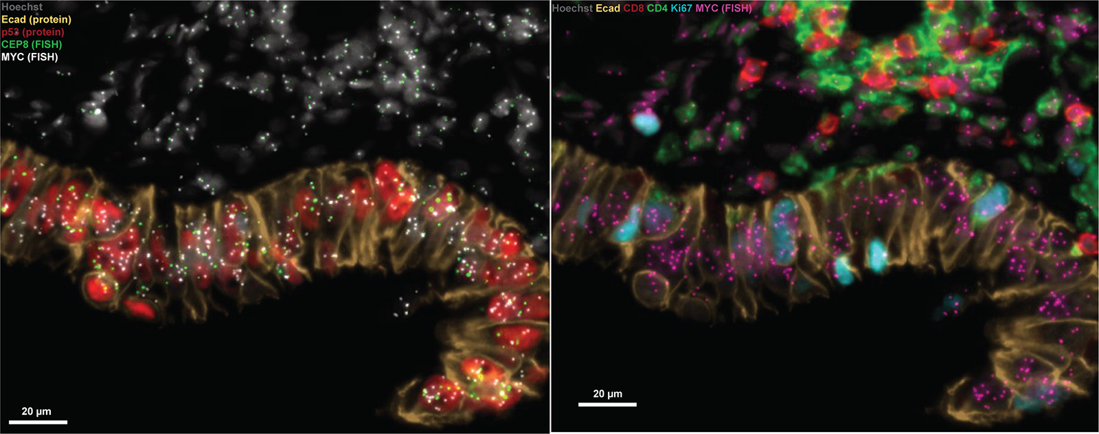

Tumors are complex ecosystems in which cancer cells carrying different somatic mutations and chromosomal copy-number changes interact with stromal and immune cells across distinctive tissue regions. Because these relationships may influence tumor behavior, preserving their geography is essential to understanding how cancers emerge and evolve. Single-cell sequencing can capture cellular heterogeneity, but it typically requires dissociation, while bulk approaches average signals across many cells. Both can obscure the spatial relationships and fine-scale interactions that are central to tumor biology. In the April issue of Cancer Discovery, Ludwig Harvard investigators Tanjina Kader and Sandro Santagata and their colleagues described ORION-FISH, a spatially resolved workflow designed to preserve that context. ORION-FISH combines multiplexed protein imaging with DNA fluorescence in situ hybridization (FISH) to map genomic alterations and the functional states and local environments of individual cells in intact tissue. Applied to high-grade serous ovarian carcinoma (HGSOC), the method recapitulated known chromosomal alterations and revealed subclonal cellular heterogeneity missed by targeted sequencing. In serous tubal intraepithelial carcinomas (STICs), precursors of HGSOC, ORION-FISH identified intermixed epithelial populations with MYC or CCNE1 copy-number gains and showed that these genomic changes were associated with distinct immune microenvironments. Some alterations were also detected in morphologically normal-appearing fallopian tube epithelial cells, providing new insight into the earliest stages of HGSOC initiation and evolution.
Spatial integration of protein and chromosomal states reveals early copy number changes and genotype-associated immune neighborhoods in serous ovarian cancer evolution
Cancer Discovery, 2026 April 18