
Dangaj Laniti lab
This image was shared by Ludwig Lausanne’s Denarda Dangaj Laniti and Fernanda Herrera.
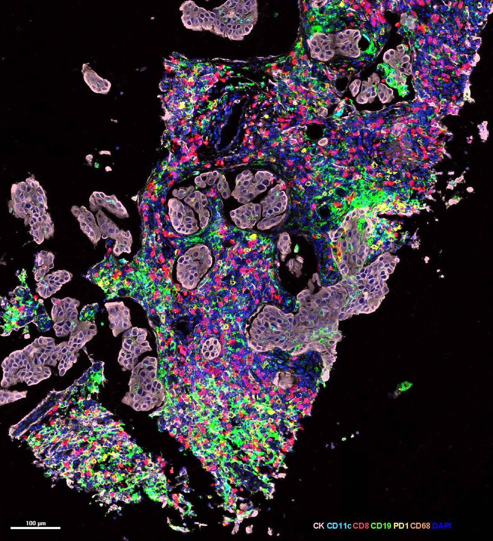
This image shows an ovarian cancer metastasis from a woman with high- grade ovarian cancer who experienced a durable complete response in the RACIN clinical trial after having recurrences following several lines of treatment. This image is from a biopsy collected ten days after low-dose radiotherapy, before anti-PD-1 checkpoint blockade had started, giving us a rare view of how radiation alone can begin to reshape the tumor microenvironment (see story).
The pale epithelial islands are cancer cells (pink). Surrounding them is a dense and colorful immune landscape, including CD8+ T cells (red), B cells (green), dendritic/myeloid cells (cyan) and other antigen-presenting cells, such as macrophages (orange). (PD-1 is stained yellow.) What makes the image powerful is the organization of these cells: immune cells are not simply present, they appear to be communicating, clustering and moving into tumor regions where anti-tumor responses can develop. For us, this image tells the story of RACIN. The study was a genuine collaboration between Fernanda Herrera’s expertise in radiation oncology and Denarda Dangaj Laniti’s work in tumor immunology and the tumor microenvironment. Together with our clinical, pathology, computational and laboratory teams, we used paired patient biopsies and deep multi-omics to ask whether low-dose radiotherapy could help convert immune-excluded tumors into lesions more receptive to immunotherapy. This image reminds us why translational research matters: it connects patient care, mechanism and discovery in a single biopsy.
Jun Nishida
This image was shared by Ludwig Harvard’s Kornelia Polyak and Jun Nishida.
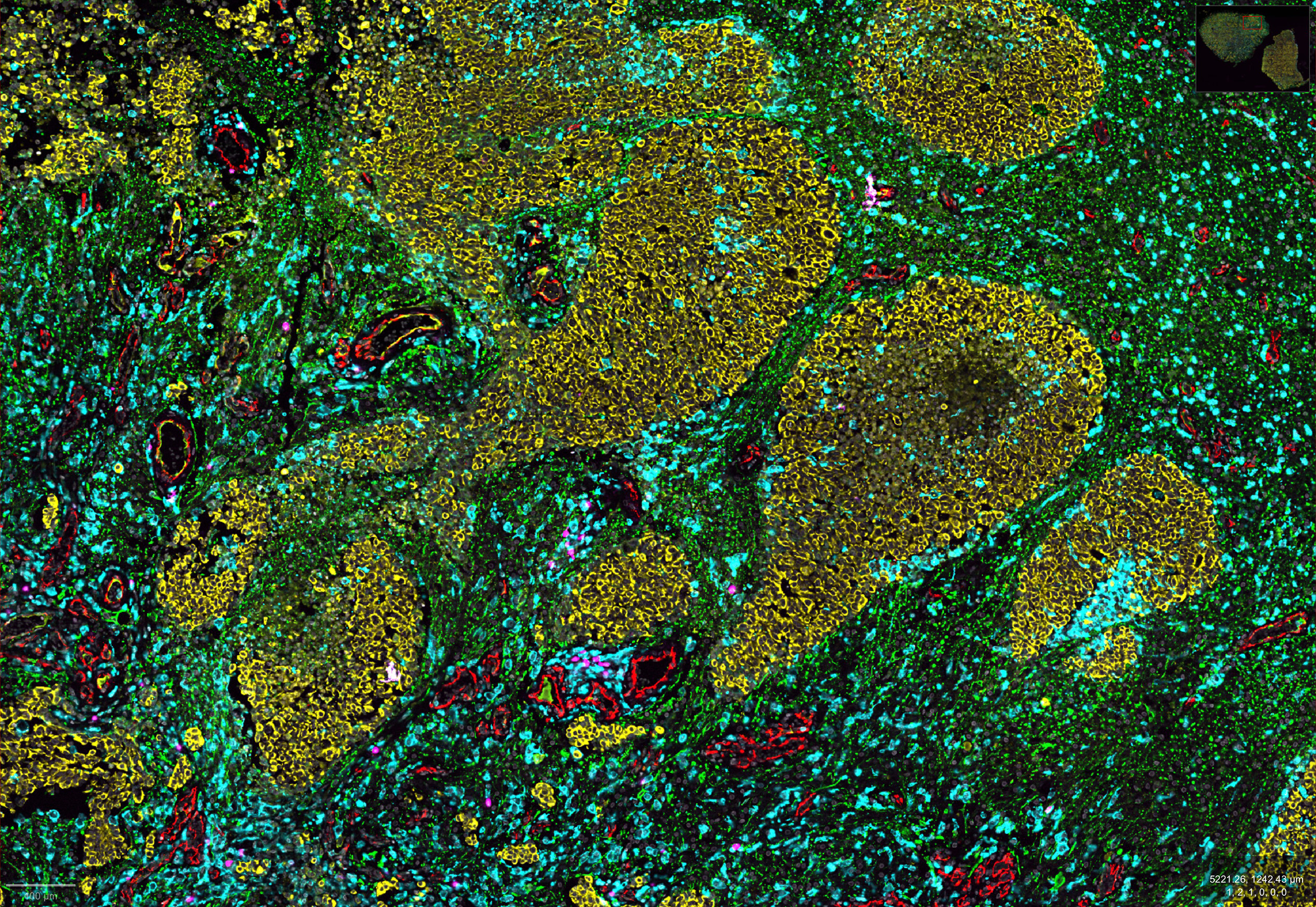
Our research aims to identify therapeutic targets of breast cancer brain metastases, with a special focus on the spatial heterogeneity of breast cancer brain metastasis and the interactions between the cancer cells and the brain microenvironment. This is a CODEX image of a breast cancer brain metastasis derived from an autopsy. The image shows a mass of invasive cancer cells (panCK+, yellow) surrounded by astrocytes (GFAP+, green) and myeloid cells (Iba1+, cyan). In these regions, very little T cell (CD3+, magenta) infiltration was observed. When seeing these images for the first time, we were amazed by the complexity and heterogeneity of the metastatic brain tumor microenvironment. The manuscript related to this image is currently under revision.
Leon Potgeter, Caroline Arber, Felix Bayerl
This image was shared by Ludwig Lausanne’s Leon Potgeter and Caroline Arber.
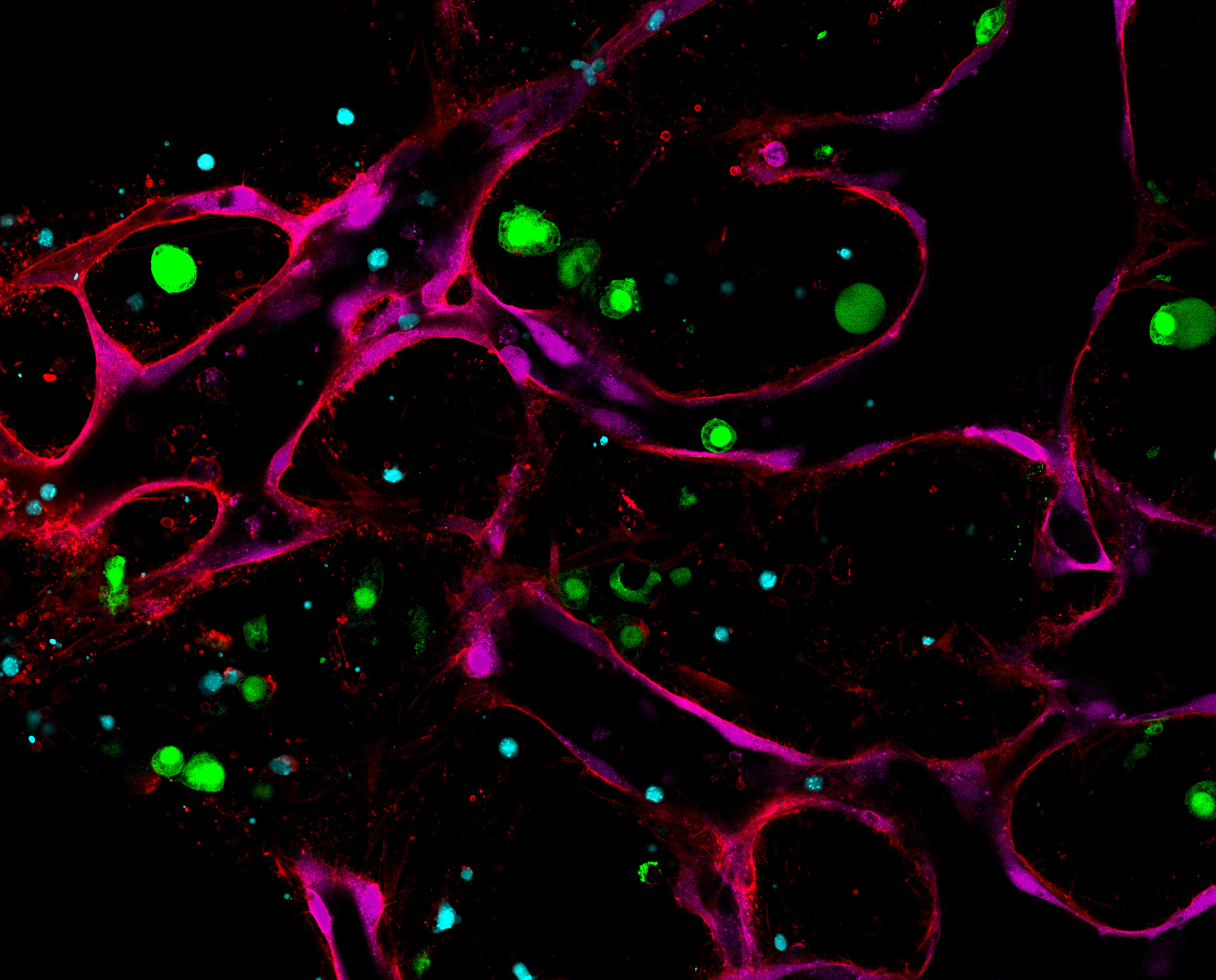
Multiple Myeloma (MM) is the second most common blood cancer. Chimeric antigen receptor (CAR)-T cell therapy has emerged as a potent treatment option for this type of cancer. However, the majority of CAR-T cell recipients eventually relapse. A major determinant of CAR-T cell efficacy is the immunosuppressive MM tumor microenvironment. This image shows a 3D in vitro MM-on-chip model that recapitulates hallmarks of the MM tumor microenvironment and is used here to investigate CAR-T cell responses against MM. MM cells (green), HUVECs—or human endothelial cells isolated from umbilical cords (magenta)—and primary human bone marrow stromal cells (CD90 stain, red) are encapsulated in a fibrin hydrogel and self-assemble into a microvascularized network supported by pericytic bone marrow stromal cells. CAR-T cells (cyan) engage MM cells (lower-left quadrant) and can be seen trafficking through the system using the engineered microvasculature. We are hopeful that this platform will support the development of novel CAR-T cell therapies, specifically therapies aimed at modulating the interplay between CAR-T cells and the MM microenvironment.
Rhea Bhalla and Avery Buchanan, Psaila lab, Khan lab
This image was shared by Ludwig Oxford’s Rhea Bhalla and Avery Buchanan.
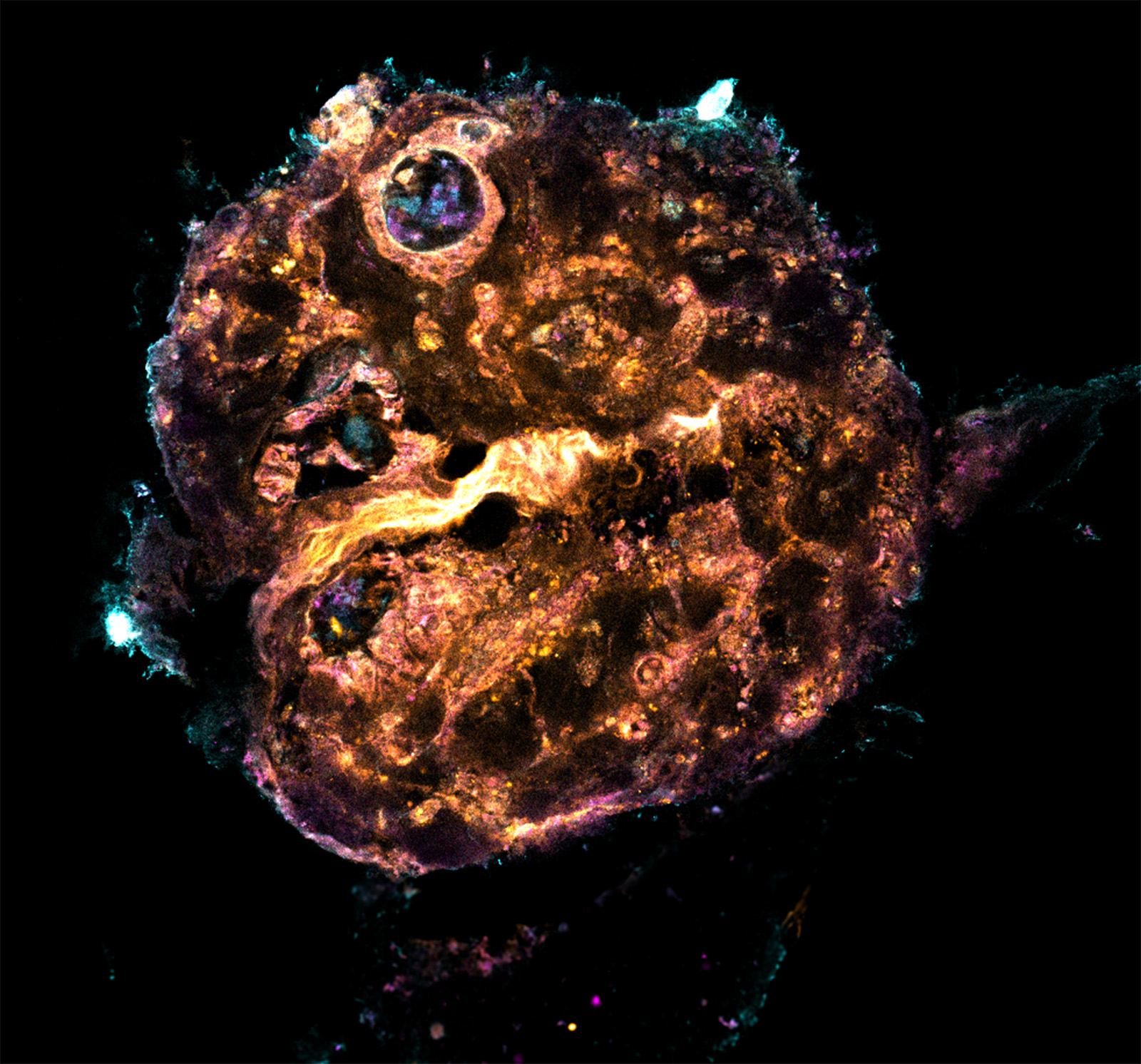
This image, which we find reminiscent of solar flares, captures a combined human bone and lympho-myeloid bone marrow organoid cultured in a rotationally and gravitationally driven, pump-free bio-compatible microfluidic device. The culture environment seeks to better replicate the bone marrow, supporting the development of a more representative in vitro tissue model. The fluidic forces and shear stress exerted on the organoid stimulate vascularization, with the aim of driving enhanced functional maturation and tissue longevity. By prolonging survival in vitro, this system enables long-term experiments and provides an improved window for studying tissue recovery following drug therapy.
Fluorescent staining highlights key cellular components—with UAE1 in orange, CD34 in magenta and CD45 in cyan—revealing the spatial organization of stromal, progenitor and hematopoietic cells. The striking distribution of these markers highlights the complex architecture that emerges within the organoid under dynamic culture conditions.
Stephen Kron
This image was shared by Ludwig Chicago’s Stephen Kron.
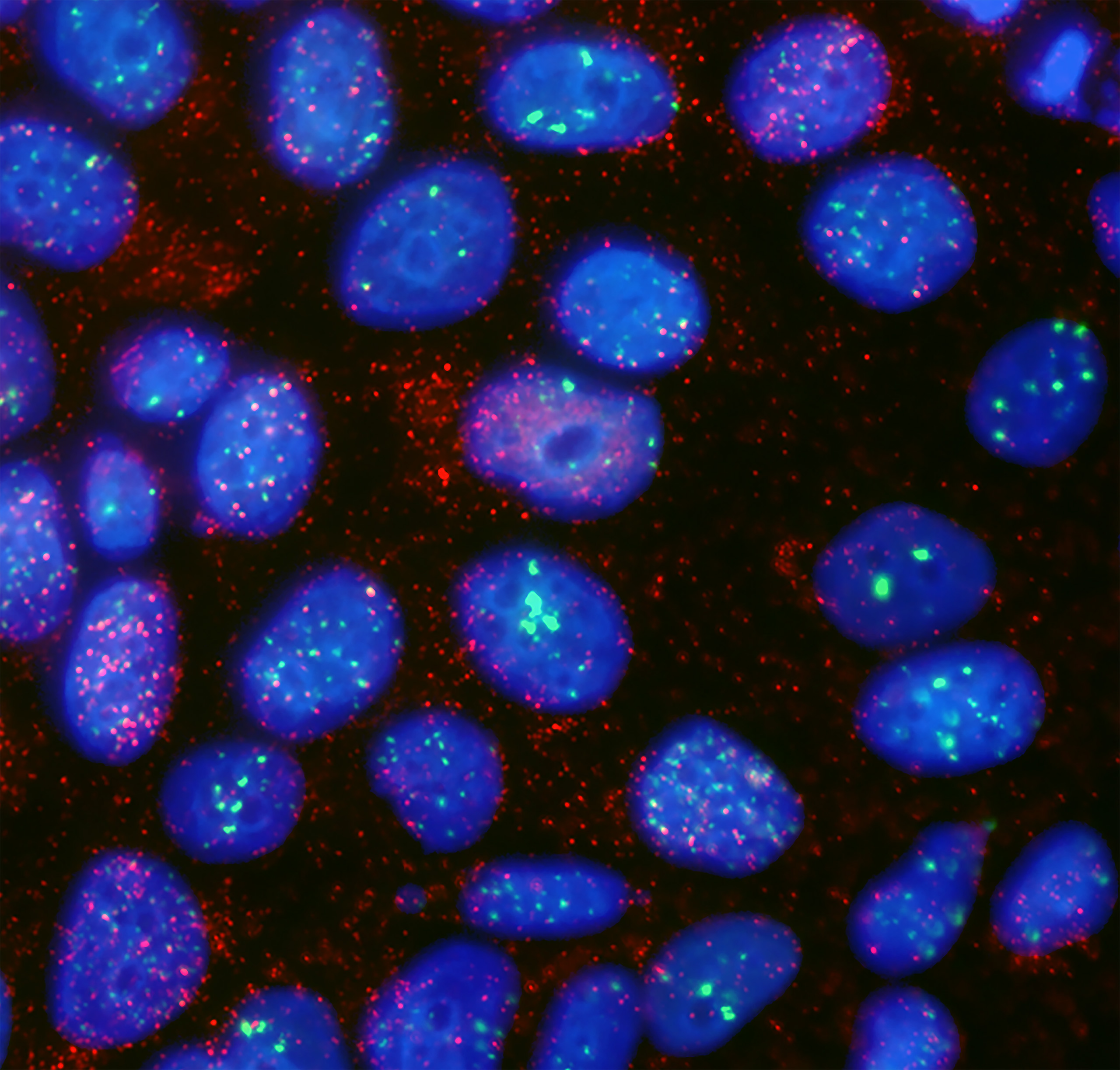
This image shows MCF7 breast cancer cells 24 hours after irradiation in the presence of the PARP inhibitor veliparib. Nuclei are stained blue, 53BP1 DNA damage foci are green, and replication protein A (RPA) bound to resected single-stranded DNA is red. The most striking feature is the appearance of RPA-positive material outside the nucleus, representing complexes of RPA with resected single-stranded DNA that have accumulated in the cytosol. PARP inhibition is well known to delay repair of radiation-induced DNA double-strand breaks, but our results suggest that something more interesting is happening. As previously reported, loss of PARP activity can promote excessive DNA end resection, or hyper-resection, producing persistent single-stranded DNA that may exceed the cell’s capacity for productive homologous recombination. What surprised us was how much of this RPA-coated DNA appeared in the cytosol, where it can engage the cGAS/STING innate immune pathway. This provides a potential mechanistic link between radiosensitization and anti-tumor immunity. PARP inhibition may not simply make DNA breaks formed in irradiated tumor cells harder to repair; it may also help convert unrepaired DNA damage into an inflammatory signal that makes the treated tumor more visible to the immune system.
Natalie Jooss
This image was shared by Ludwig Oxford’s Natalie Jooss.
I am a postdoc in Beth Psaila’s group at Ludwig Oxford, where I investigate blood cancers known as myeloproliferative neoplasms (MPNs). My research focuses on the role of the novel biomarker Galectin-1 and its underlying mechanisms in disease progression within the bone marrow niche as well as its effects on circulating platelets.
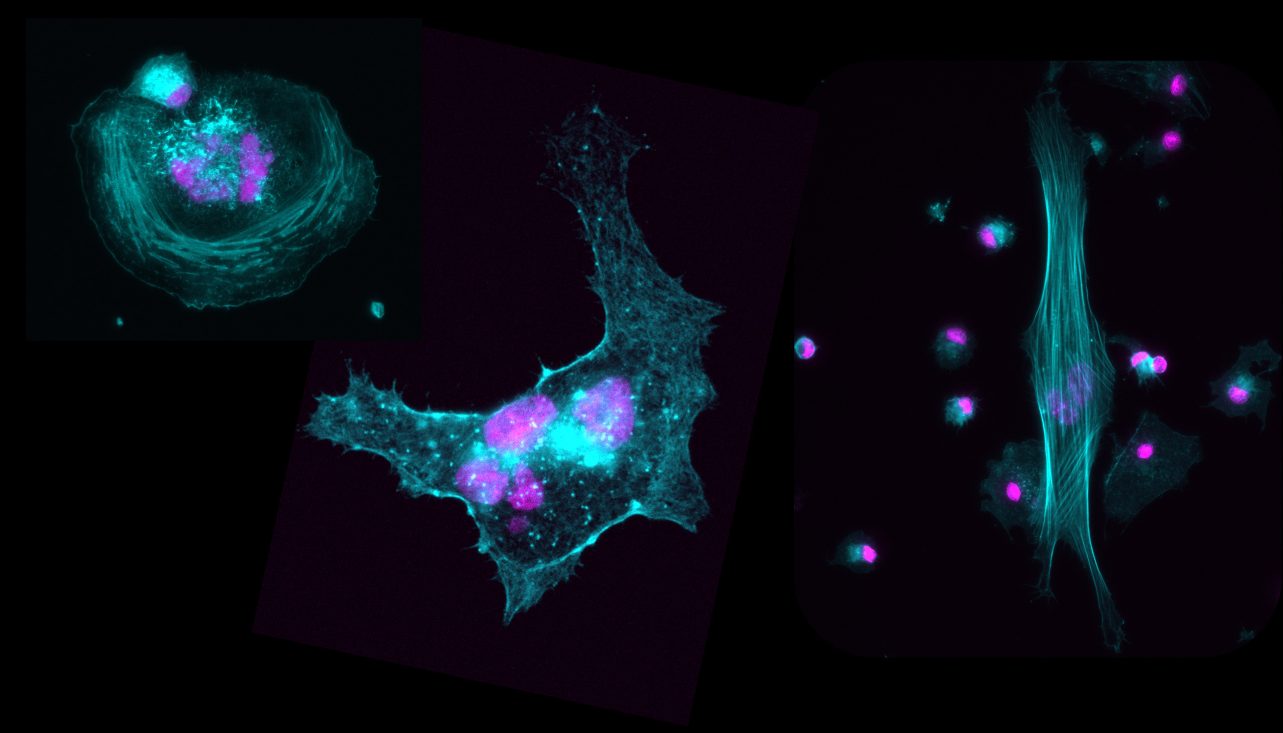
This image comes from our investigations into how changes in the bone marrow microenvironment affect megakaryocytes (Mks), the precursors of platelets.
Mks, generated here from induced pluripotent stem cells (iPSCs), are remarkable cells. As they mature, they become much larger than most other cells and often develop polyploid nuclei, making them particularly impressive to observe under a microscope. Here, fluorescence microscopy reveals the cells’ cytoskeleton in cyan and their nuclei in magenta. What struck me most was the diversity of the cells. Although they originated from the same iPSC line each developed its own distinctive shape.
That element of surprise is one of the things I find most exciting about science. Microscopy allows us to visualize a world that is normally completely hidden from us, and experiments intended to answer scientific questions can unexpectedly reveal just how intricate and beautiful biology can be.
Benita Wolf, UNIL/CHUV (cells: Jandus lab)
This image was shared by Benita Wolf (Department of Oncology, UNIL/CHUV), in collaboration with Ludwig Lausanne’s Camilla Jandus.
My group studies how cytotoxic T cells engage and kill tumor cells at the nanoscale. To do this, we adapted cryo-expansion microscopy (cryo-ExM) for T cell biology: cells are frozen at −180 °C, embedded in a swellable hydrogel and physically enlarged, so that structures well below the diffraction limit become resolvable on a conventional confocal microscope. The technique is particularly suited to visualizing the immunological synapse.
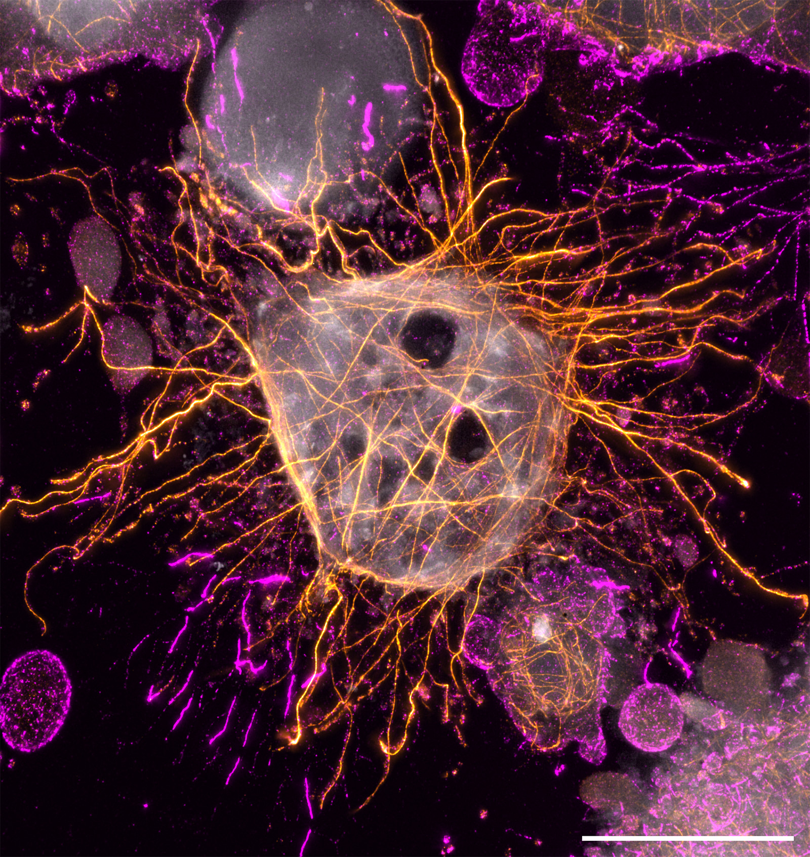
Displayed here is a dying human melanoma cell (GEFI) after co-culture with cytotoxic CD4+ T cells, labeled for actin (magenta) and tubulin (orange), with an NHS-ester counterstain of primary amines (gray). Scale bar: 10 μm (corrected for the expansion factor). The cells were provided by Camilla Jandus’s lab; sample preparation, cryo-ExM and imaging were done in my lab.
As it happens, this image never made it into our study, because there is no bona fide immunological synapse in it, which is what we were looking for. But we keep coming back to it. What surprises us is the microtubule cytoskeleton in this dying cell: the way the microtubules still organize around the nucleus at a point when the plasma membrane has already lost its integrity. Cell death is usually described as a collapse, and here something is very clearly still organized. Whether that organization means anything, or whether it is simply what a cytoskeleton looks like on the way down, is a question we can’t answer from a single image.