Puzzlers and drug designers
For more than a century, researchers have parsed the structure and biochemistry of the centrosome. A study led by Ludwig’s Karen Oegema, Andrew Shiau and Timothy Gahman finally solved two lingering mysteries about the structure, and may have opened the door to a new strategy for treating cancer.
Peering into fertilized roundworm eggs in the late 19th century, the legendary German zoologist Theodor Boveri identified a minute, beadlike structure associated with spindles that yanked chromosomes apart in the dividing cells of the embryos. Boveri named this “special organ of cell division” the centrosome and, over the next three decades or so, never quite took his eye off the structure. In 1914, a year before his death, he formally hypothesized that multiple copies of centrosomes fueled malignancy by inducing the accumulation of abnormal numbers of chromosomes in cancer cells.
Since then, researchers have parsed the structure and biochemistry of centrosomes and elucidated their many vital contributions to the cell’s inner life. But it has remained unclear whether rapidly dividing cancer cells are indeed “addicted” to multiple copies of centrosomes. Ditto, oddly enough, for whether they’re even needed for the proliferation of normal cells. In 2015, a full century and a year after Boveri published his hypothesis, a collaborative study led by Ludwig San Diego’s Karen Oegema, and Andrew Shiau and Timothy Gahman of the Small Molecule Discovery Program, finally answered both questions. Their paper, published in Science, may have opened the door to implementing a strategy for treating cancer fielded some two decades ago by Ludwig’s scientific director, David Lane.
THE PUZZLERS
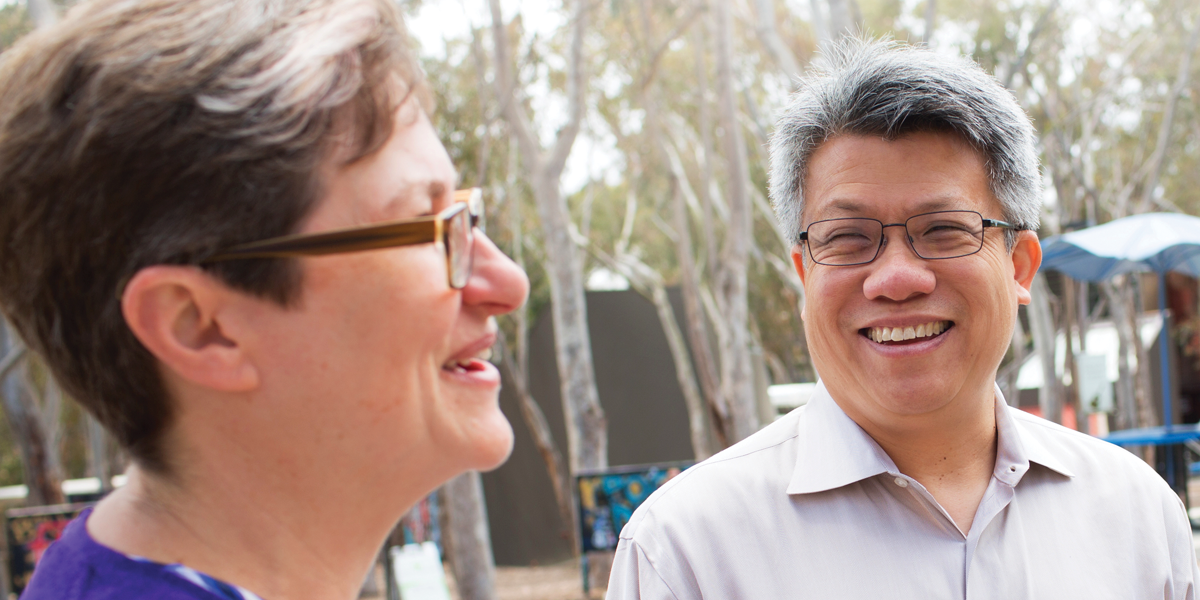
When Oegema talks about the cell, or all of the things she can do with her microscopes, she sounds a lot like a kid in a candy store. Only, the treats in her case are some of the central problems of cell biology. “I have commitment issues,” she jokes. “We wind up working on a lot of different projects in my lab.”
Today, these include probing how dividing cells split in two, and using a spectacular, advanced high-content microscopy system obtained by Ludwig to chart out, on a grand scale in the flatworm C. elegans, what it is that the proteins encoded by all the genes involved in cell division and embryonic development exactly do.
But like Boveri, Oegema has a bit of a soft spot for the centrosome, whose biochemistry she studied for her doctoral research in the mid-1990s at the University of California, San Francisco (UCSF). “Centrosomes,” she recalls, “were held up as a sort of Holy Grail in cell biology. It was very difficult to crack what their components do, define the scope of their role in cells and to describe precisely how they duplicate.”
Oegema’s Science paper stemmed from her work dissecting centrosome duplication in C. elegans—echoed by discoveries others had made in mammalian cells—which suggested that a protein known as Plk4 controls the assembly of centrioles, barrel-like structures from which centrosomes are made. Oegema was interested in blocking this protein as a route to asking a range of basic biological questions.
Enter Shiau, who has known Oegema and her husband, Ludwig’s Arshad Desai, since graduate school at UCSF and now heads Ludwig’s Small Molecule Discovery Program. He and Gahman, who is a medicinal chemist, both worked for years in the biotech industry. They have a wealth of experience in the design and development of drug-like molecules for research and therapy and support Ludwig researchers around the world in this capacity.
“We look for problems like this, where investigators have a committed interest in a pathway or a problem,” says Shiau. “Then Tim and I ask, is there a fundamental problem in cancer biology that can be answered?”
TEAMING UP
Oegema’s proposal, they thought, fit the bill. “The centrosome question was very much on people’s minds,” says Oegema. “For a hundred years people have known that cancer cells have too many. The question is, why? Are they some sort of driving force in the genesis of tumors?”
The cell’s single resident centrosome serves as an organizing center for its cytoskeleton, an intricate network of protein filaments that, among other things, confer shape, internal organization and motility upon cells. When a cell divides, though, the centrosome takes on its most famous function. It duplicates and helps ensure the equal distribution of chromosomes to the two daughter cells.
Biologists have, however, long known that other mechanisms exist to pull chromosomes apart, and many researchers once believed that centrosomes may not be required for cell division. On the other hand, it was clear that multiple centrosomes do contribute to the misdistribution of chromosomes seen in cancer cells.
To solve these mysteries, researchers had long sought to remove centrosomes by surgically excising them from cells or blasting them with lasers. But both normal and cancer cells treated this way simply remade their lost centrosomes and then continued dividing. Groups in academia and industry have tried to develop Plk4 inhibitors to target centrosomes. These work in the test tube—shutting Plk4 down—but, for a variety of reasons, couldn’t be used to stop cells from remaking their centrosomes.
To get around this limitation, the researchers designed and then tested hundreds of inhibitors for their effects on centrosomes in living cells. “We didn’t care if we had a powerful inhibitor of Plk4 in the test tube,” says Shiau. “What we cared about was getting a molecule that does what we want it to do inside the cell. You get what you screen for.” It took a while, but the team finally came up with a molecule that specifically and—this was crucial to the subsequent study—reversibly inhibited Plk4.
They showed that exposure to the molecule, which they named centrinone, eliminates centrosomes from both healthy and cancerous cells. Normal cells treated with centrinone simply stopped dividing and went into a state of latency known as arrest. Cancer cells, on the other hand, did not stop dividing when their centrosomes were removed, though fewer survived the ordeal.
“What we learned using centrinone is that normal cells do really care about having centrosomes and have the ability to detect their loss,” says Oegema. Conversely, cancer cells are not addicted to multiple centrosomes. In fact, they continue dividing, albeit less efficiently, even when they have none.
HISTORY, AGAIN
The researchers showed that the pause in the division of healthy cells is governed by a tumor suppressor named p53. Dubbed the Guardian of the Genome, p53 is mutated in about half of all cancers. A couple of decades ago, Lane—a co-discoverer of p53—suggested that its role in stopping cell division in response to trouble of all sorts might be exploited for cancer therapy.
“The idea,” says Shiau, “is that you trigger p53 in normal cells and have them stop multiplying—and then introduce another agent that only kills continuously dividing cells.” Though centrinone is not a drug, molecules related to it may enable the experimental assessment of Lane’s idea, which he named cyclotherapy. The new ability to reversibly eliminate centrosomes is also likely to benefit research in a wide variety of biomedical fields, given the organelle’s multiple roles, from organizing the cytoskeleton to sprouting hair-like structures known as cilia on certain cells.
“Centrinone is a very good research tool, and we have been giving it out freely to academics and nonprofits,” says Gahman. “Our next step is to ask what centrosome removal will do in animal models of cancer, and so we’ve made better, more drug-like molecules. It’s one thing to look in a dish and see centrosomes go away. It can be quite a different thing when you’re in a live tumor, in a living animal. That’s where we’re going now.”