The cancer cell cuisinologist
After making landmark contributions to our understanding of how cancer cells evade programmed death, Eileen White discovered that they also depend on a process of self-cannibalization, known as autophagy, to survive. Her work is elucidating its profound influence on tumor biology and opening exciting new possibilities for cancer therapy.
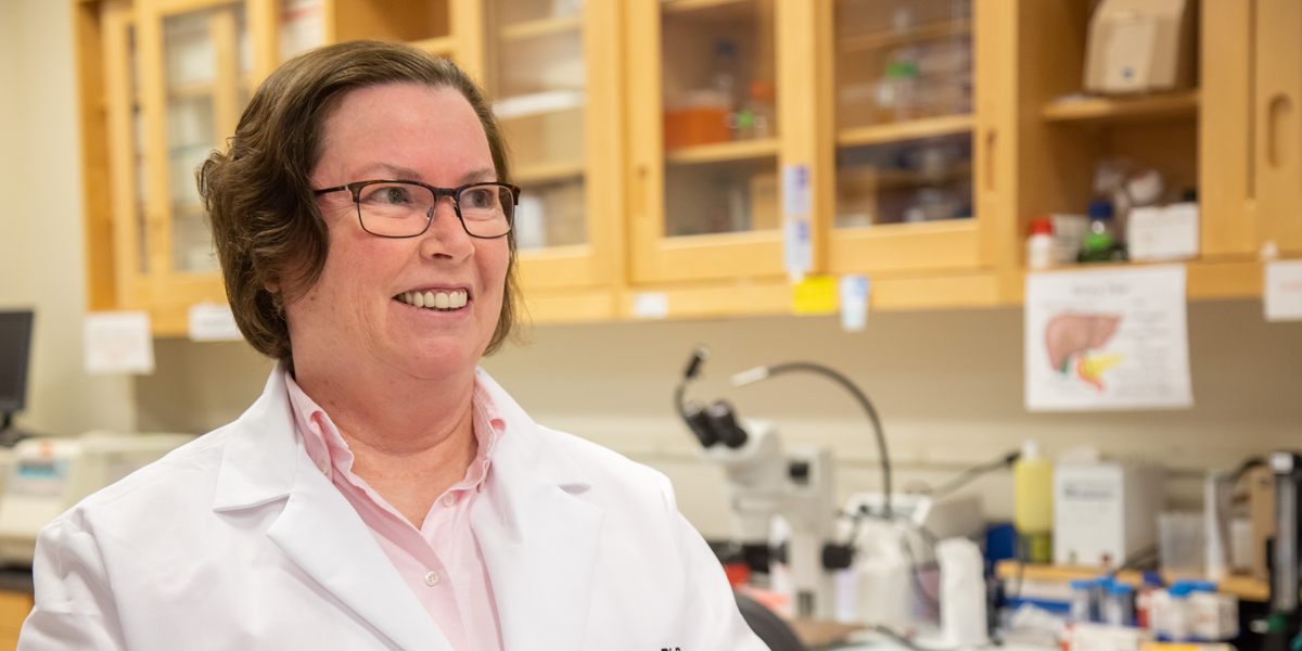
Eileen White wasn’t quite sure what she wanted to do for her postdoctoral research as she wrapped up her graduate studies at the Stony Brook campus of the State University of New York. But she was sure about a couple of things: she wanted to take on something really difficult, and she wanted to make it count. As she mulled over how to do that, White learned of an opening in Bruce Stillman’s group at the famed Cold Spring Harbor Laboratory nearby on Long Island. Stillman was interested in how a set of oncogenes identified in the recently sequenced genome of the adenovirus caused cancer. That focus—at the intersection of cancer research and virology—perfectly fit her criteria, and White joined Stillman’s group in 1983.
It was a fortuitous choice and a perfect fit for a young researcher with a yen for big problems. “Bruce said, ‘This is the virus, this is the oncogene, figure out how it causes cancer’,” recalls White, who is today a distinguished professor of molecular biology and biochemistry at Rutgers University in New Jersey and associate director of Ludwig Princeton. “To me, that was a gift. What a great project for a postdoc!”
Her work on that adenoviral oncogene played a key role in establishing the importance of apoptosis, or programmed cell death, in suppressing cancer, and helped launch a field of cancer research that has already yielded new therapies targeting apoptosis blockers and promises to generate many more. That research, in turn, led to White’s landmark identification of autophagy—in which cells cannibalize themselves to recycle nutrients—as an important survival mechanism of cancer cells, launching yet another subfield of cancer research with notable promise for the development of new therapies. It has also drawn White deep into the study of cancer metabolism, the focus of the Ludwig Princeton Branch.
WAIT A MINUTE!
The daughter of a lawyer and an elementary school teacher, White grew up in a small town on Long Island, New York. Her father had as a teenager hoped to become a physicist and retained a fascination with science. His influence contributed to White’s interest in biology, which she majored in as an undergraduate at the Rensselaer Polytechnic Institute in New York, before pursuing graduate studies in Eugene Katz’s laboratory at the State University of New York, Stony Brook, studying the genetics of development.
Not quite cancer research, but viral oncology was then in its heyday, and Stony Brook’s Department of Microbiology was at the forefront of the field. Arnold Levine, the departmental chair, had recently isolated in complex with a viral antigen the cellular p53 tumor suppressor protein, whose function is now known to be disrupted in more than half of all cancers. The department also counted among its faculty at the time Ludwig Harvard Co-director Joan Brugge, who had previously discovered and characterized a viral oncogene named Src and shown that the human genome encodes its homologue—a milestone of modern cancer research. Both were mentors to White, who received her PhD in 1983.
At the time, it was almost an article of faith that viral oncogenes function exclusively by promoting cell proliferation, the fundamental, unifying characteristic of cancers. Another principle taking shape in the field—one that has survived the test of time—was that multiple oncogenic mutations are required to initiate cancer. White’s work would help confirm the latter concept, while debunking the former.
The adenoviral gene Stillman assigned to White was named E1B 19K. Its oncogenic partner in the viral genome was named E1A. White was told that only when inserted into a healthy cell together—not individually—would the gene pair transform it into a cancer cell. But in repeating those experiments White noticed that when E1A alone was inserted into cells, colonies of proliferative cells would indeed form. Puzzled, she reported this observation to more experienced lab members.
“They said to me, and this is a direct quote, ‘Don’t worry, those colonies will go away’,” White recalls. “I said, ‘what do you mean, they’ll go away?’ But it was true: the colonies formed, and then the cells died. Then, when I put E1A and E1B in together, the colonies didn’t die. That’s when the light bulb switched on in my head. This suggested to me that E1A was the one driving proliferation, and E1B was allowing the colonies to survive. This was conceptually novel at the time.”
White suspected that E1B was supporting survival by preventing apoptosis, or programed cell death, a phenomenon that was poorly understood at the time. As it happened, the Harvard scientist Stanley Korsmeyer had discovered that an oncogene in cells, Bcl-2, drove cancer by preventing apoptosis. White believed E1B was the viral equivalent of Bcl-2, so she contacted Korsmeyer and proposed that they collaborate to test that hypothesis. In studies that continued after White had set up her own lab at Rutgers in 1990, the pair demonstrated that this was indeed the case.
White’s group subsequently reported that the suppressed apoptosis is mediated by the p53 protein and contributed key discoveries on how p53 induces cell suicide. They and other researchers would also demonstrate that the Bcl-2 family includes proteins that either drive or suppress apoptosis. Whether or not a cell commits suicide depends on the balance of these opposed proteins. “What we—the whole field—wanted to do ultimately was get a therapy that inhibited the anti-apoptotic proteins to unleash the pro-apoptotic proteins for cancer therapy,” White explains.
They succeeded: new drugs that tip the balance in favor of apoptosis are now approved or in the late stages of development for cancer therapy. White’s contributions also helped establish the suppression of apoptosis as a hallmark of cancer and underscored p53’s critically important role as a tumor suppressor.
“In the beginning, when your cells died, you threw them in the garbage,” White says. “I was one of those people saying, wait a minute…!”
All this while, White had also remained busy on the administrative front. Soon after she arrived at Rutgers, the university had recruited William Hait from Yale University to set up and direct a new cancer center. “Bill had an enormous task at hand,” says White. “He had to build a cancer center from scratch. I mean, there was nothing.”
Hait asked White to join the effort, and with her help obtained a Comprehensive Cancer Center designation from the National Cancer Institute. Today, White is the chief scientific officer and deputy director of the Rutgers Cancer Institute of New Jersey. The Ludwig Princeton Branch will be conducting many of its translational clinical studies in partnership with the Institute.
STUBBORN SURVIVORS
By the late 1990s, White’s lab was brimming with cancer cell lines engineered to be defective in apoptosis.
“We were doing all sorts of things to cells to find out what it meant to be unkillable by apoptosis, and there were things we couldn’t understand,” says White. “You put them in buffer and went home for the weekend, and they’d still be alive. It didn’t make any sense. Making a cell unable to kill itself should not impart a miraculous ability to survive starvation.”
Hoping to discover their secret, White and her colleagues put the cells under an electron microscope and noticed they were packed to the proverbial gills with autophagosomes. These are small membranous sacks typically containing the cell’s defunct proteins and old organelles, destined for dismantling and recycling. Though mammalian autophagy was not well understood at the time, a vast body of work had established its genetics and metabolic chemistry in yeast, which switch on autophagy during starvation. White and her team thought that their never-say-die cancer cells might be doing the same thing.
It turned out, however, that the cancer cells gobbled up their innards even when they were swimming in nutrients. White’s subsequent studies examining this unexpected discovery demonstrated for the first time that autophagy is a key mechanism of survival for solid tumor cells.
“Autophagy is important when you’re starved or stressed—your cells turn it on, stuff gets recycled, and you’re fine,” says White. “But what we were seeing was that cancer cells were usurping that pathway. Anytime you find a survival pathway that cancer cells are using, you have to block it. No one in my lab wanted to work on anything else.”
The White lab’s experiments established, first, that autophagy is a survival mechanism: knocking out the genes essential to autophagy killed cancer cells, especially when they were simultaneously starved or subjected to other types of stresses.
“The other function of autophagy is quality control, getting rid of the garbage—bad organelles, bad protein aggregates, things that are toxic,” White explains. “So the two fundamental properties that it impinged on were metabolism and inflammation, because when you don’t throw out the garbage, that triggers inflammation.” Inflammation, meanwhile, is an invitation to immune attack, which tumors take great pains to avoid.
WADING INTO METABOLISM
By the late 2000s, White’s lab began working extensively with genetically engineered mouse models of cancer to examine how autophagy influences tumor metabolism. To get a better handle on the new discipline, White began a close and continuing collaboration with Josh Rabinowitz, who helped pioneer the field of metabolomics—the large-scale, quantitative analysis of metabolites—and is now director of Ludwig Princeton.
“Josh is down the road from us at Princeton, and is the master of all things metabolism,” says White. “It’s really been wonderful because when we first began working together, he did not know very much about cancer and I did not know much about metabolism. We’ve learned a lot from each other. It is a perfect example of scientific synergy.”
Over the next decade and a half, White’s lab would demonstrate in mouse models the importance of autophagy to lung, prostate, breast and melanoma tumors. Using lung cancer models, for example, they showed that disabling autophagy at the point of cancer initiation results in tumors with lower proliferation and malignancy and a higher level of apoptosis compared to controls.
They also discovered, in an early collaboration with Rabinowitz’s group, that in cancer cells autophagy is critical to ensuring a steady supply of amino acids to mitochondria—the bean-like organelles that generate cellular energy. That supply ensures the optimal function of the TCA cycle, a core series of metabolic reactions essential to mitochondrial energy generation. TCA cycle dysfunction depletes reservoirs of molecules required to make DNA and other cellular components required for cell proliferation. “Knock out autophagy in these cells,” says White, “and they suffer a metabolic crisis.”
In another series of studies, White and Rabinowitz compared in an animal model of lung cancer how autophagy loss in cancer cells alone affects tumor growth compared to its systemic loss in the mouse. They found that cancer cells die far more quickly than healthy ones following the loss of autophagy, suggesting that the strategy might well be safely and effectively employed for therapy. In fact, tumors regressed far more dramatically when autophagy was inhibited systemically and didn’t grow at all if it was already absent.
This suggested that the autophagy conducted by healthy cells also supports tumor growth. Their studies revealed that mice deficient in autophagy had low circulating levels of an amino acid, arginine, that drives tumor cell proliferation. It turned out that the livers of autophagy-deficient mice were secreting an enzyme that breaks down arginine, an essential tumor nutrient; dietary arginine could, they showed, partially revive tumor growth.
INTO TUMOR IMMUNOLOGY
But that wasn’t the only reason tumors weren’t thriving in autophagy-deficient mice. Previous studies done by White, Rabinowitz and their colleagues had shown that autophagy reshapes the global expression of proteins in cancer cells to eliminate proteins that drive inflammation and can provoke a lethal immune response. White’s group had also found that in mice deficient in autophagy, antibodies that removed T cells—which kill cancerous and infected cells—could restore tumor growth to some degree.
In 2020, White led a study in collaboration with several other labs showing that systemic autophagy suppresses the anti-cancer immune response. In its absence, cancer cells activate the frontline cells of the innate immune system, initiating a cascade of molecular events that culminates in the recruitment and activation of anti-tumor T cells.
“By inactivating autophagy, we were unleashing inflammation in a way that activates T cells to kill tumor cells,” says White. “A whole flurry of papers that came out around the same time found that autophagy profoundly suppresses the anti-tumor immune response.”
White and her colleagues also discovered that autophagy in the liver is most responsible for that inhibition. This opens up the possibility that preferentially targeting autophagy in the liver in combination with other treatments could improve outcomes of cancer therapies and immunotherapies. White’s lab is now conducting studies to better understand the phenomenon.
With cancer metabolism and immunology now major areas of study in her lab, it’s little surprise she was excited to join Ludwig Princeton. “It’s not like Josh had to twist my arm,” White says, recalling how she reacted to being recruited to the Branch. “It was an amazing opportunity, everything all wrapped up together. It was perfect.”
White’s membership in Ludwig connects her to an accomplished community of tumor immunologists and an institution that helped launch the modern era of cancer immunotherapy. “Being a part of the Ludwig Princeton Branch will allow Josh and me to expand our collaboration to new areas to achieve even greater impact. We are very excited to tackle mechanisms of cancer metastasis with Ludwig Princeton’s Yibin Kang and to build on success in cancer immunology with other Ludwig researchers.”
Immunotherapies, White says, are among the most remarkable achievements of cancer research, but they need to be made applicable to a far broader range of cancers. She suspects the manipulation of cancer metabolism could be key to that effort.
“This is the perfect time to target metabolism in cancer,” says White. “There are already anti-metabolites in our armamentarium of cancer drugs, but now we can understand metabolism at a level we never previously could. We can now more effectively take advantage of seeing cancer as a metabolic disease.”